Cracked Tooth Syndrome is characterized by fine cracks that can extend from the crown all the way down to the root of the tooth and are often not visible to the naked eye. Symptoms tend to fluctuate. Sometimes there is no pain for days, and sometimes a single bite can trigger a sharp, stabbing pain. For this reason, it is a challenging condition to diagnose for both the patient and the dentist. It disrupts chewing patterns and can progress to tooth loss. In this article, we’ll clearly summarize the symptoms of Cracked Tooth Syndrome, the tests we use, and realistic treatment options.
What Is Cracked Tooth Syndrome?
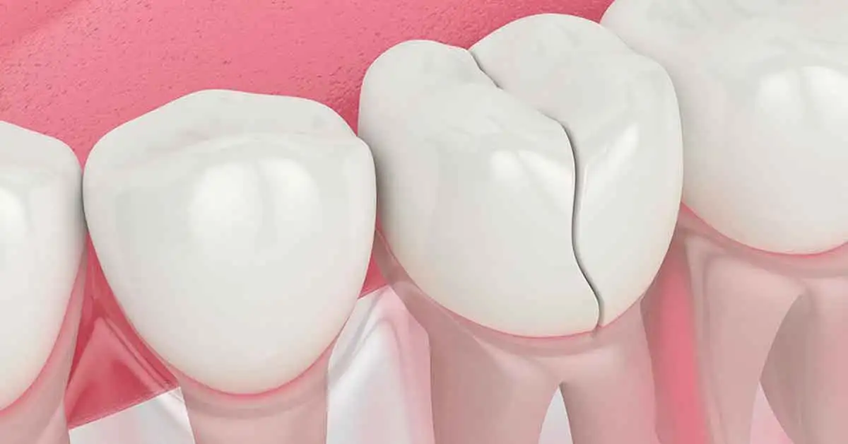
Cracked Tooth Syndrome is characterized by fine fracture lines in the enamel and dentin of the tooth, and sometimes extending down toward the root. There is no fragmentation like in a complete fracture. The tooth remains in the mouth. However, with each bite, the crack line moves microscopically. This movement delivers mechanical and hot–cold stimuli to the nerve.
We most frequently see it in molar teeth. Habitual consumption of hard foods, teeth grinding or clenching, old large fillings, and root canal–treated teeth are important risk factors. If a cracked tooth is not treated, it can eventually split into two separate pieces. In that case, the chance of saving the tooth decreases.
What Are the Symptoms of Cracked Tooth Syndrome?
Symptoms of Cracked Tooth Syndrome are usually fluctuating. Most patients describe it as, “Nothing shows up on the X-ray, but when I chew it feels like a knife stabbing inside the tooth.” Common findings include:
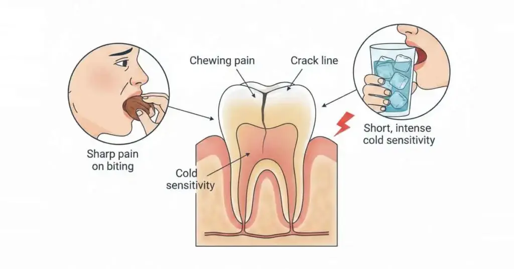
• Sharp, needle-like pain when chewing on a specific point
• Sensitivity to hot and cold (especially cold)
• A stabbing pain in the tooth when eating something sweet
• Pain when biting down, with relief when releasing the bite
• Some days no complaint at all, other days severe pain
• Avoiding chewing on the same tooth or side when pressure triggers pain
Not every case of Cracked Tooth Syndrome presents the same way. Sometimes there is only mild discomfort when chewing hard foods. Sometimes it progresses to pain that wakes the patient up at night. The gums often look normal. This can mask the underlying problem.
Risk Factors for Cracked Tooth Syndrome
Although Cracked Tooth Syndrome often feels like it appeared suddenly, there is usually a cumulative load built up over many years. The most common risk factors we see are:
• Teeth grinding and clenching (bruxism)
• Habitual consumption of hard foods (nuts, roasted chickpeas, seeds, ice chewing)
• Large, old metal or composite fillings
• Root canal–treated teeth with significant loss of tooth structure
• Trauma (blows, accidents, sudden excessive force on the tooth)
• Unbalanced bite, excessive load concentrated on certain teeth
Patients in these groups have a significantly higher risk for Cracked Tooth Syndrome. Molars with very large fillings that have been in use for many years should be followed very closely.
How Is Cracked Tooth Syndrome Diagnosed? (Clinical Tests)
Cracked Tooth Syndrome is not diagnosed with a single X-ray. The first step is taking a detailed history. We ask which bite, on which side, and what type of pain is felt. Then we perform clinical and radiographic tests:
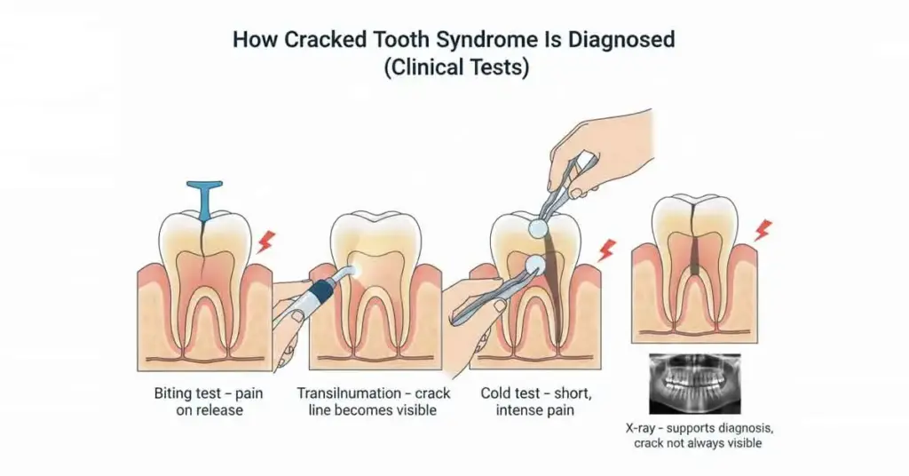
• Bite tests:
We use special bite blocks or cotton rolls. The patient slowly bites down with the suspected tooth and then releases suddenly. Pain is typically felt most at the moment of release. This finding is characteristic of Cracked Tooth Syndrome.
• Percussion test:
We gently tap on the tooth to check for tenderness. If the crack has progressed, pain may increase with vertical tapping.
• Cold test:
We apply a controlled cold stimulus and observe the tooth’s response. Prolonged and intense pain can indicate that the crack is affecting the pulp.
• Transillumination (light examination):
We shine a strong light source onto the tooth. The crack line appears as a dark band where the light is interrupted.
• Examination under magnification:
We examine the tooth surface in detail using loupes or a microscope. Enamel cracks especially become more apparent this way.
• Radiographs and 3D imaging when needed:
Standard X-rays do not always show the crack itself. However, we always take them to rule out surrounding bone loss, root fractures, and other pathologies. If needed, we use CBCT (3D tomography) to check for fractures at the root level.
By combining these tests, we get closer to a diagnosis of Cracked Tooth Syndrome. The goal is to understand both the direction and the depth of the crack.
Treatment Options for Cracked Tooth Syndrome
Treatment for Cracked Tooth Syndrome is planned according to the location and depth of the crack. Not every cracked tooth has to end in extraction. The main approaches we use include:
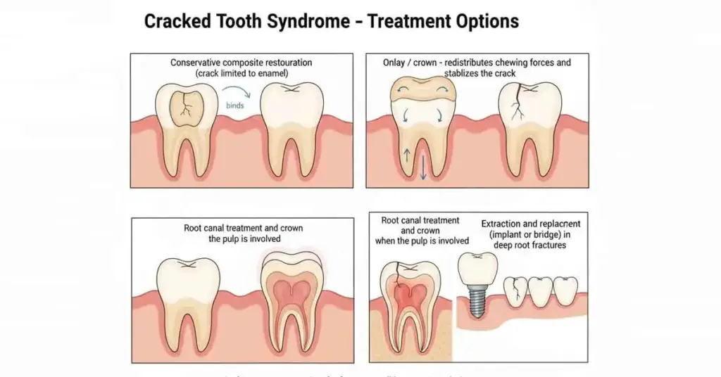
• Conservative repair and composite restoration:
If the crack is limited to the enamel, we remove the weakened tissue and reshape the tooth as a single unit with composite filling material.
• Reinforcement with onlay/crown:
If the crack has extended into the dentin and the tooth walls have become thin, we plan a crown or onlay that will wrap around the tooth “as one piece.” The aim is to distribute chewing forces over the surface and stop movement along the crack.
• Root canal treatment:
If the crack has affected the pulp and long-lasting pain with hot/cold has begun, root canal treatment becomes necessary. After root canal treatment, we always reinforce the tooth with a crown. In these teeth with significant loss of structure, Cracked Tooth Syndrome can be triggered again if not properly protected.
• Extraction and implant/bridge planning:
If the crack extends all the way to the root and a vertical root fracture has occurred, the chance of saving the tooth is low. In such cases, the tooth is extracted. A dental implant or bridge is then used to replace the missing tooth. Here, the aim is to preserve both chewing function and bone volume.
In every Cracked Tooth Syndrome case, the first goal is to keep the existing tooth in the mouth and in function. However, when deciding, we evaluate the X-ray findings, the clinical picture, and the patient’s habits together.
How to Protect a Tooth With Cracked Tooth Syndrome
Once we diagnose Cracked Tooth Syndrome, there are important steps the patient should take as well:
• Avoid hard foods (raw almonds, ice, very hard or shell-on foods)
• Avoid chewing only on one side, on the same area
• Wear a night guard if there is teeth grinding or clenching
• Have the treated tooth checked regularly
• Protect gum health with interdental cleaning and regular brushing
These behaviors slow the progression of the crack and extend the lifespan of the treatment. Once Cracked Tooth Syndrome has developed, that tooth is considered a “high-risk tooth.” That’s why periodic follow-up is extremely valuable.
“Cracked Tooth Syndrome is one of the most common reasons behind the sentence, ‘There’s nothing on the X-ray, but my tooth still hurts.’ With early diagnosis and proper treatment, it is possible to save the tooth before it reaches the point of extraction. If you feel sudden, pinpoint pain while chewing, the safest approach is not to dismiss it as ‘just temporary sensitivity,’ but to see a dentist in a timely manner.”
Cracked Tooth Syndrome – FAQ
Does Cracked Tooth Syndrome heal on its own?
No. There is no mechanism where the crack closes and the pain disappears. The crack line makes micro-movements with every bite. As long as this movement continues, the nerve is stimulated. Fluctuating pain does not mean it is healing.
Does a tooth with Cracked Tooth Syndrome always have to be extracted?
Not always. If the crack is limited to the enamel and outer dentin, we can often keep the tooth in the mouth for many years with appropriate fillings and a crown. Extraction is more common in vertical fractures that extend to the root.
Nothing shows up on my X-ray. Could it still be Cracked Tooth Syndrome?
Yes. Standard dental X-rays often do not show fine cracks. Diagnosis is made through clinical tests, patient history, and examination under magnification. Cracked Tooth Syndrome can be “invisible” on radiographs.
Is root canal treatment always necessary for Cracked Tooth Syndrome?
Not in every case. If the crack has not progressed into the pulp chamber, the tooth is still vital, and the pain is short-lived, conservative restoration may be enough. If the pulp is affected, then root canal treatment comes into play.
Can Cracked Tooth Syndrome come back?
Even after a crack is repaired, that tooth has already carried heavy loads in the past. If teeth grinding continues, a night guard is not used, or excessive force is applied with hard foods, new cracks may develop. That’s why changing habits is an essential part of treatment.